That said, knowing what to look for makes a real difference. This article covers what facial moles actually are, what causes them, how to distinguish a benign mole from one that warrants a dermatologist visit, and the clinically approved removal options available today.
Key Takeaways
- Most adults develop 10 to 40 moles by adulthood; the majority are benign
- Use the ABCDE rule to self-check for asymmetry, irregular borders, colour variation, large diameter, and any evolution over time
- Use the ABCDE rule to self-check moles for warning signs at home
- A mole appearing after age 40 or changing rapidly deserves prompt evaluation
- Clinically approved removal methods (shave excision, surgical excision, RF cautery) are always safer than DIY attempts
- Daily SPF 50+ sunscreen is your most effective tool for limiting UV-driven mole darkening and new formation
What Are Facial Moles? Types and Appearance
A facial mole — clinically called a melanocytic nevus — is a benign cluster of melanocytes, the pigment-producing cells normally distributed throughout your skin. When these cells group together rather than spreading evenly, they form the small dark spots we recognise as moles. According to the American Academy of Dermatology, most adults with lighter skin develop between 10 and 40 common moles, with many appearing during childhood and adolescence.
The Three Types Worth Knowing
| Type | Key Characteristics |
|---|---|
| Common Acquired Nevi | Appear after birth; round or oval, evenly coloured, usually under 6mm; may be flat or raised |
| Congenital Melanocytic Nevi | Present at birth; occurs in roughly 1 in 100 people; size ranges from small to large/giant |
| Dysplastic (Atypical) Nevi | Larger than typical moles, irregular or notched border, mixed colouring — not melanoma, but warrants monitoring |
Moles vs. Similar-Looking Spots
Not every dark spot on your face is a mole. Dermatosis papulosa nigra (DPN), for example, presents as multiple small pigmented papules common in people with darker skin tones. It's actually a variant of seborrheic keratosis — an epidermal growth, not a melanocytic one. Seborrheic keratoses can look strikingly similar to moles but respond to entirely different treatments. Getting a dermatologist's assessment before pursuing any removal option ensures you're treating the right thing.
What Causes Moles on Your Face?
Moles form when melanocytes — the cells responsible for skin pigment — cluster in one spot rather than spreading evenly. Several factors drive that clustering:
- Genetics: A family history of many moles or atypical nevi raises your likelihood of developing them. Both mole count and characteristics tend to run in families.
- UV exposure: Intermittent sun exposure and childhood sunburns are linked to higher acquired mole counts. UV radiation stimulates melanocyte activity, which can darken existing moles or trigger new ones over time.
- Hormonal shifts: Puberty and pregnancy are commonly associated with new mole formation or visible changes in existing ones. However, one study tracking 129 nevi prospectively found little mean size change during pregnancy — hormonal fluctuations alone are rarely cause for concern.
When Mole Count Becomes a Risk Factor
Most facial moles are completely benign. However, certain markers increase the risk of melanoma:
- 50 or more moles anywhere on the body
- Four or more atypical moles, especially with a personal or family history of melanoma
- Large or giant congenital nevi present from birth
These are risk indicators, not diagnoses — but they do mean regular monitoring becomes more important.
Symptoms of Facial Moles: How to Identify Normal vs. Concerning
What a Normal Mole Looks Like
A benign facial mole typically has:
- Round or oval shape with symmetrical halves
- Uniform brown or tan colouring throughout
- Smooth or slightly raised texture
- Diameter under 6mm (roughly the size of a pencil eraser)
- Stability over months and years — no new symptoms
The ABCDE Rule
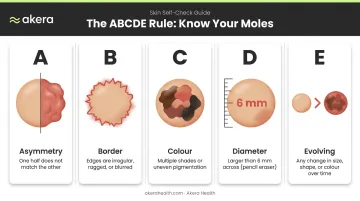
The ABCDE criteria, introduced by Friedman, Rigel, and Kopf and later updated to include evolution, remain the gold-standard self-check tool:
- A — Asymmetry: One half doesn't match the other
- B — Border: Irregular, notched, scalloped, or blurred edges
- C — Colour: Multiple shades within one mole — tan, brown, black, red, or white
- D — Diameter: Larger than 6mm (though melanomas can be smaller)
- E — Evolving: Any change in size, shape, colour, or new symptoms like itching or bleeding
The Ugly Duckling Rule
The Ugly Duckling sign — a lesion that looks distinctly different from all your other moles — is a helpful second check alongside the ABCDE criteria. A 2017 study found a specificity of 0.96 for clinical images; comparing moles against each other reduced unnecessary biopsies by nearly 7-fold. The practical takeaway: if one mole stands out as the odd one, get it checked.
Two Things Most People Get Wrong About Moles
Elevation alone tells you nothing. A mole can be flat or raised based purely on its growth pattern — what actually matters is asymmetry, border irregularity, colour variation, and change.
New adult moles aren't automatically cause for alarm either. Mole count typically increases until your 30s or 40s. A mole appearing for the first time after 40 that grows or changes is the one that warrants attention.
Red-flag symptoms that need prompt evaluation:
- Spontaneous bleeding from a mole
- Persistent itching or pain
- Rapid changes over just a few weeks
- A mole that crusts or fails to heal
When Should You See a Dermatologist for a Facial Mole?
See a dermatologist if any ABCDE criterion applies, if a mole has visibly changed in recent months, or if a new spot appears after age 40 and seems to be growing. Early evaluation is always the safer call — localized melanoma has a 5-year relative survival rate of 100% according to U.S. SEER data (2016-2022), compared to 34% for distant-stage disease.
What to Expect at a Dermatologist Appointment
A dermatologist evaluation typically involves:
- Visual assessment — including dermoscopy in many cases, which uses magnification and light to examine the mole's structure in detail
- History review — how long the mole has been present, whether it's changed, and any family history of melanoma
- Biopsy if needed — if there's any suspicion of malignancy, a small tissue sample is taken for analysis
One persistent concern is worth addressing directly: biopsying a mole does not cause cancer to spread. The NCI and American Cancer Society both describe biopsy-related seeding as extremely rare, and the diagnostic benefit far outweighs the theoretical risk.
How Often Should You Check?
- Monthly self-skin exams — familiarise yourself with your moles so you notice changes quickly
- Annual professional skin exam — or more frequently if you have a family history of melanoma, many moles, or atypical nevi
If you're in Bengaluru, Akera Health (HSR Layout and HRBR Layout) offers dermatologist-led skin assessments under Dr. Lavina Mittal (MBBS, MD) — covering mole evaluation, dermoscopy where indicated, and a clear discussion of whether any follow-up is needed.
Facial Mole Removal: Options and What to Expect
People seek mole removal for two legitimate reasons: medical necessity (suspicious moles, or moles that cause physical irritation) and cosmetic preference. Both are valid, and both start with the same step — a dermatologist evaluation.
Clinically Approved Removal Methods
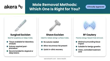
Surgical excision cuts out the mole along with a margin of surrounding healthy skin, then closes the wound with sutures. Dermatologists prefer it for suspicious or deeper moles because it provides tissue for histological examination and carries the lowest recurrence risk. The trade-off is a slightly longer scar line.
Shave excision removes a raised mole flush with the skin surface without sutures. It's well-suited for benign, elevated moles. A 2021 randomised trial of 145 nevi found that shave excision produced smaller scars and higher patient satisfaction compared to elliptical excision, though recurrence occurred in 11.7% of shave cases — exclusively from shave technique — due to residual cells left in the dermis.
RF cautery (radiofrequency cautery) uses high-frequency energy to precisely remove benign skin growths with minimal impact on surrounding tissue. At Akera Health, this is one of the confirmed mole removal methods offered alongside shaving and excision.
A note on laser removal: dermatological consensus advises against laser mole removal as a primary treatment because no tissue specimen remains for histological analysis, and recurrence with altered appearances has been documented.
The Scarring Reality
Your choice of removal method directly shapes the scar outcome. Modern minimally invasive techniques performed by a trained dermatologist keep marks minimal, and proper aftercare matters just as much as the technique itself.
Never attempt DIY removal. The AAD warns that at-home methods — apple cider vinegar, garlic, online tools — carry serious risks: infection, uncontrolled bleeding, permanent scarring, and the very real danger of missing a cancerous mole. A documented case report described a chemical burn following apple cider vinegar nevus removal.
Most procedures are outpatient treatments done under local anaesthesia, taking only minutes to complete. At Akera Health, mole removal is performed under dermatologist supervision with treatment plans tailored to your skin type, mole characteristics, and cosmetic goals — so you leave with a clear aftercare plan and realistic expectations for healing.
Aftercare and Preventing New Facial Moles
Post-Removal Care
Following mole removal, the AAD recommends:
- Wash hands before touching the area; gently clean with mild, fragrance-free soap and water
- Apply petroleum jelly (not routine antibiotic ointment, which can cause allergic reactions) and cover with a sterile dressing
- Avoid pools, hot tubs, and prolonged water exposure until the area heals
- Protect the healing site from direct sun exposure — UV on a fresh wound increases the risk of hyperpigmentation and visible scarring
- Attend all follow-up appointments as scheduled
Limiting New Mole Formation
You can't prevent moles entirely, but reducing UV exposure is the most effective way to limit new ones and prevent existing moles from darkening. Once healing is underway, consistent sun protection becomes your primary long-term defence.
The WHO advises limiting midday sun exposure between 10am and 4pm, particularly when the UV Index reaches 3 or higher — a threshold that India's cities regularly exceed.
Practical steps:
- Apply broad-spectrum SPF 50+ sunscreen daily — including on overcast days — and reapply every two hours outdoors
- Wear wide-brimmed hats and UV-protective clothing when outdoors
- Establish a monthly self-skin check routine and schedule annual dermatologist visits
For post-procedure skin specifically, a non-comedogenic SPF 50+ formulated for your skin type is worth prioritising. Akera Health's sunscreen range covers oily/combination, normal, and dry/sensitive skin — the dry/sensitive variant is particularly suited to healing or reactive skin.
Frequently Asked Questions
Can moles be removed from the face?
Yes. Facial moles can be safely removed using shave excision, surgical excision, or RF cautery, among other methods. The right approach depends on the mole's size, depth, and clinical status, and should always be decided in consultation with a qualified dermatologist. At Akera Health, mole removal is performed by certified dermatologists to minimise risk and ensure clean results.
What is the cause of moles on the face?
Facial moles form when melanocytes cluster together rather than distributing evenly across the skin. Genetics, UV sun exposure, and hormonal changes during puberty or pregnancy all contribute to their development, number, and tendency to darken.
How do I stop moles from appearing on my face?
You can't prevent moles entirely, but daily broad-spectrum SPF 30+ sunscreen, protective clothing, and avoiding peak UV hours (10am–4pm) help reduce both new mole formation and darkening of existing ones.
How do I know if my facial mole is cancerous?
Use the ABCDE rule — Asymmetry, irregular Border, uneven Colour, Diameter over 6mm, and Evolution — as a starting point. Any mole showing these signs needs professional evaluation; only a dermatologist examination, and potentially a biopsy, can confirm whether it is benign or malignant.
Does facial mole removal leave a scar?
Some scarring is possible with any method, but minimally invasive techniques performed by a trained dermatologist keep marks small. Consistent aftercare — sun protection, gentle cleansing, and moisturising — helps the skin heal with minimal visible scarring.